


ORIGINAL ARTICLES
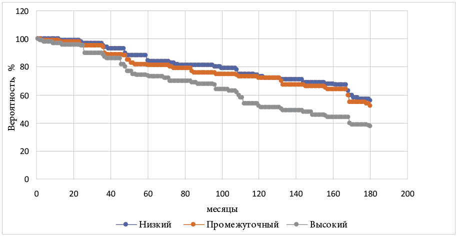
Introduction. Prostate cancer (PC) is considered to be one of the most common malignancies in men. In recent years, conventional PC treatments have been supplemented with highly effective minimally invasive therapies such as highintensity focused ultrasound ablation (HIFU). Only a few studies have been published on the long-term oncological effectiveness of HIFU therapy for prostate cancer. Aim. To evaluate the oncological efficacy of HIFU in the treatment of prostate cancer in a long-term follow-up. Materials and methods. The retrospective analysis included the treatment outcomes of 171 patients with prostate cancer who underwent HIFU therapy at the Samara Clinical Oncology Dispensary in 2007–2009. The mean age of the patients was 69.7 years. Of these, 48 had a low risk of progression according to D’Amico, 57 patients — intermediate risk and 66 — high risk of progression. The follow-up period comprised 13–15 years (median 14.3 years). Positive histological findings, elevated PSA and/or the appearance of local or distant metastases were interpreted as recurrence. The Kaplan-Meier method was used to graphically represent survival curves. A multiparameter Cox proportional hazards regression analysis was performed to assess the prognostic significance of various clinical data in overall, cancer-specific and recurrence-free survival. All values of p<0.05 were considered statistically significant. Results and discussion. The overall fifteen-year survival for patients in the low-risk, intermediate-risk, and high-risk groups accounted for 52.1, 56.1, and 37.9%, respectively. Fifteen-year PC-specific survival was determined in 90.1% of patients. Fifteen-year recurrence-free survival for patients in the low-risk, intermediate-risk, and high-risk groups comprised 95.4, 80.7 and 69.7%, respectively. A significant risk factor for recurrence was the distribution according to the D’Amico progression risk scale. Conclusion. HIFU therapy demonstrated good long-term oncologic results in the treatment of patients with prostate cancer.
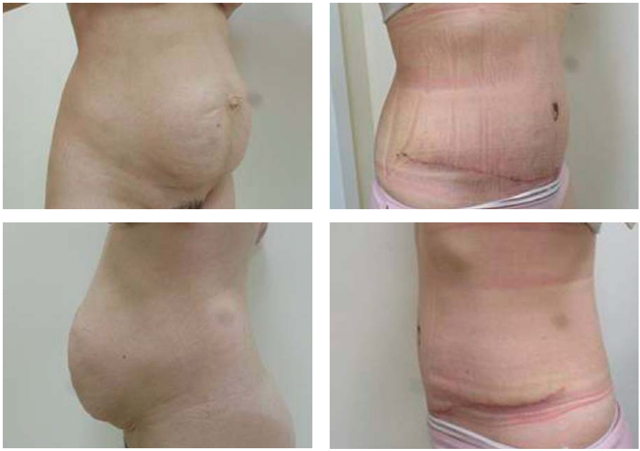
Introduction. According to the estimates of many countries, liposuction comprises a sought-after aesthetic surgery, achieving a leading position among popular surgical interventions in recent decades. For instance, the records of the American Society of Plastic Surgeons show, that liposuction occupied the 4th place in 2020, 3rd — in 2019, and 2nd — in 2000. Liposuction is frequently included in other aesthetic surgeries. In 2019–2023 the Plastic Surgery Unit of the Mother and Child Clinical Hospital, Ufa, performed this surgical intervention an isolated operation, and as part of abdominoplasty, mammoplasty, brachioplasty, and face-lifting. Aim. To analyze the clinical data of the Plastic Surgery Unit of the Mother and Child Clinical Hospital, Ufa, on the experience of performing liposuction as a stage of the anterior abdominal wall correction in isolated abdominoplasty and simultaneous hernioabdominoplasty. Materials and methods. The study involved 56 patients: women with overweight or obesity, suffering from aesthetic and functional problems in the anterior abdominal wall associated with pregnancy and childbirth. Surgical treatment and outpatient follow-up were performed in the Plastic Surgery Unit of the Mother and Child Clinical Hospital, Ufa from 2019 to 2023. Group 1 of patients included 33 people who underwent abdominoplasty combined with liposuction, group 2 comprised 23 patients, who had hernioabdominoplasty with liposuction. Results and discussion. Liposuction refers to a technique for removing excess fat deposits and improving the contours of the anterior abdominal wall. Liposuction stands as the main component of surgical intervention on the anterior abdominal wall, simplifying the stages of detachment of the anterior abdominal wall and redistribution of the skin-fat flap. The complication rates in abdominoplasty with liposuction and hernioabdominoplasty with liposuction differ insignificantly. Conclusion. Liposuction as a part of abdominoplasty or hernioabdominoplasty contributes to the improvement of surgical treatment outcomes.
Introduction. Idiopathic pulmonary fibrosis (IPF) comprises an interstitial lung disease with unclear pathogenesis, rapid progression, and no effective treatment. Pulmonary fibrosis is reported to be one of the most severe complications induced by a new coronavirus infection COVID-19. The mechanisms triggering pulmonary fibrosis and leading to its rapid progression remain substantially unclear. Evidence suggests that immune and genetic factors contribute to the development of this disease. Among the latter, the role of long non-coding RNAs (dnRNAs) has been actively studied to date. Materials and methods. Considering the role of TP53TG1, LINC00342, H19, MALAT1, DNM3OS, and MEG3 dnRNAs as regulators of signaling pathways associated with fibroblast activation and epithelial-mesenchymal transition, the authors analyzed the expression level of selected dnRNAs in lung tissue and blood mononuclear cells of patients with IPF (N = 12), post-COVID-19 pulmonary fibrosis (N = 14), and in control group (N = 27). Results and discussion. Blood mononuclear cells in patients with IPF and post-COVID-19 PF revealed similar patterns of TP53TG1 and MALAT1 dnRNA expression. The level of relative expression of MALAT1 was significantly higher in patients with IPF (Fold Change=3.207, P = 0.0005) and with post-COVID-19 PF (Fold Change=9.854, P = 0.0003), while the relative expression level of TP53TG1 reduced in patients with IPF (Fold Change=0.4308, P = 0.0313) and with post-COVID-19 PF (Fold Change=0.1888, P = 0.0003 in blood mononuclear cells, Fold Change=0.1791, P = 0.0237 in lung tissue). Increased expression of DNM3OS in blood mononuclear cells (Fold Change=12.899, P = 0.0016) and lung tissue (Fold Change=9.527, P = 0.0001), LINC00342 (Fold Change=2.221, P = 0.0309) in blood mononuclear cells was revealed only in patients with IPF. Conclusion. Evaluation of the dnRNA expression profile of TP53TG1, LINC00342, MALAT1 and DNM3OS in blood mononuclei can be used as an informative and non-invasive biomarker in IPF and post COVID-19 PF.
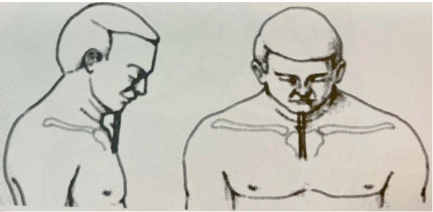
Introduction. Cicatricial tracheal stenosis comprises a severe complication of traumatic or prolonged intubation of the trachea. Circular resection of the trachea serves as a radical method of surgical treatment. The distension of the anastamosis can be prevented by immobilization of the neck and head in the thoracic adduction, as a rule, with the Grillo suture technique. This method of fixation creates severe discomfort for the patient, especially when eating. Rare cases involve neurological disorders due to compression of the neurovascular bundles of the neck and spinal cord. Materials and methods. The study enrolled 6 patients diagnosed with cicatricial tracheal stenosis upon admission to the thoracic surgery unit. All patients underwent a circular tracheal resection. Immobilization of the cervical spine was performed using a cervical collar (Schantz collar type). Results. Immobilization was performed within 7–13 days, followed by a control tracheobronchoscopy. No complications related to anastomotic failure and no specific complications when using these orthoses were revealed. Discussion. The postoperative period was reported as uneventful; anastomotic healing proceeded in accordance with the physiological timing indicated by the framework of other studies. The applied orthosis provides immobilization of the cervical spine, preventing from any movement in this area, which is actually similar to the use of the Mulliken-Grillo suture, but has a number of advantages: absence of cosmetic defects, possibility of quick fixation and removal. Significantly, this provides an opportunity to assess the risk of neurochemical damage in patients during preliminary fixation of the head in the hyperflexic position at the preoperative stage. Conclusion. Having such advantages as the absence of cosmetic defects and the possibility of quick removal, this method of neck fixation can be considered as an alternative to the classical fixation method.
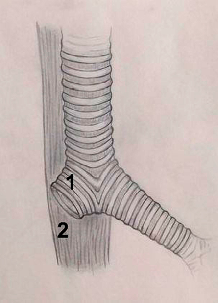
Introduction. At present, bronchial stump failure after pneumonectomy ranges from 1% to 20%, and mortality — from 20 to 75%. In past decades, various methods of preventive bronchoplasty have been introduced and improved in order to reduce the incidence of the bronchial stump failure and bronchopleural fistula. However, the existing methods fail to prove their predominant effectiveness to be suggested as a “gold standard” for prevention of the bronchial stump failure during pneumonectomy. A method introduced by the authors enables an additional tissue mobilization with uncontrolled circulatory disturbance of the “biological flap” to be avoided. Aim. To evaluate the results and effectiveness of preventive esophagomyobronchoplasty in clinical practice. Materials and methods. The suture of “preventive esophagomyobronchoplasty” of the bronchial stump after pneumonectomy in lung cancer was developed and introduced into the clinical practice of the Thoracic Surgery Unit. The study involves a statistical analysis of bronchial stump failures as well as bronchopleural fistulas before and after the introduction of preventive esophagomyobronchoplasty. Results and discussion. A retrospective analysis of 224 clinical observations proves a zero probability of such complications after pneumonectomy as bronchial stump failure and bronchial fistula in case of preventive esophagomyobronchoplasty. The incidence of pleural empyema does not exceed 1.3%. Notably, pleural empyema implies longer duration of stay, additional examination, expensive drugs, as well as direct threatening life of a patient. Conclusion. Since 2015, when the new method of bronchial stump formation has been introduced into clinical practice, the rate of complications in the form of bronchial stump failure was reduced from 10.5% to 0%.
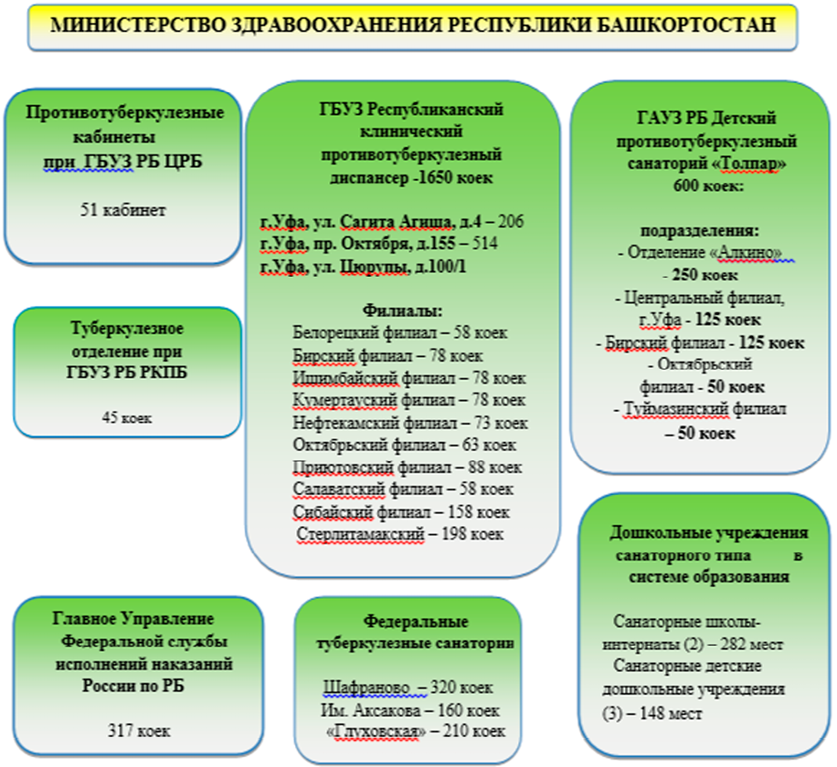
Introduction. Surgical intervention is recognized as an integral part of pulmonary tuberculosis treatment. Optimal routing of pulmonary tuberculosis patients to surgical treatment essentially improves the effectiveness of tuberculosis treatment. The paper is aimed at analyzing the routing of pulmonary tuberculosis patients to surgical treatment in the Republic of Bashkortostan. Materials and methods. The paper presents the structure of Republican Clinical Tuberculosis Dispensary, describes the routing of patients followed by the TB Service of the Republic of Bashkortostan, indicates a significant role of telehealth technologies in providing specialized medical care for patients with pulmonary tuberculosis. In addition, the paper introduces the quantitative data and structure of surgical interventions performed in the TB Surgery Unit of the Republican Clinical Tuberculosis Dispensary. Results and discussion. According to the conducted analysis, the TB Service of the Republic of Bashkortostan complies with the regulatory documents of the Russian Federation, thus ensuring the most complete coverage of pulmonary tuberculosis patients in need of thoracic surgery. Conclusion. Timely routing of patients to a TB surgery unit enables effective treatment, differential diagnosis, and abacillation of patients to be provided, thereby reducing the spread of tuberculosis in the region.
REVIEWS

Organs-on-chips (OOC) refer to microfluidic devices used to create biomimetic systems of physiological organs. The system contains engineered or natural miniature tissues grown inside microfluidic chips. Organ-on-a-chip technology enables numerous human pathologies to be reproduced, since classical animal models may fail to adequately predict the therapeutic response in humans. This technology can be an intermediate link in the animal-human research system. Moreover, in cancer studies, OOC simulate the three-dimensional hierarchical complexity of tumors in vivo and the tumor microenvironment, being an efficient and cost-effective solution for tumor growth studies and cancer drug screening. Organs-on-chips represent compact and easy-to-use microphysiological functional units simulating physical and biological processes in human body. This extends the possibility of preclinical studies, such as disease modeling or even the development of diagnostic devices. In this regard, the present study is aimed at reviewing the scientific literature in the field of microfluidic devices intended for use in urology and oncourology.
C ervical cancer remains a pressing global health problem, creating a significant health burden for women worldwide. High incidence and mortality rates necessitate further research to unravel its underlying molecular mechanisms and identify new diagnostic and treatment strategies. Recent advances in non-coding RNAs have opened up new avenues for research, including circular RNAs (circRNAs) as molecules that play a multifaceted role in cellular processes. Research into circRNAs revealed their unique structure, characterized by the covalent formation of a closed loop, thereby distinguishing them from their linear counterparts. These circRNAs are involved in regulating various aspects of cell physiology with a particular focus on cell growth and development. Interestingly, circRNAs have context-dependent functions, acting both as promoters and inhibitors of oncogenic processes, depending on the complex cellular environment in which they operate. Recent studies have identified aberrant expression patterns of circRNAs in the context of cervical cancer, implying their key role in the disease development. The different expression profiles of circRNAs associated with cervical cancer offer promising opportunities for early detection, accurate prognosis assessment, and personalized treatment strategies. The presented comprehensive review offers an in-depth study of cervical cancer-associated circRNAs, their specific functions and complex molecular mechanisms driving the onset and progression of cervical cancer. Increasing evidence suggests that circRNAs can serve as invaluable biomarkers for early detection of cervical cancer and promising therapeutic targets for intervention. Delving into the complex interaction between circRNAs and cervical cancer paves the way for innovative and personalized approaches to combat this serious disease, aiming at reducing its impact on women’s health worldwide and improve patient outcomes. Unraveling the mysteries of circRNAs in the context of cervical cancer makes the prospects for a breakthrough in its diagnosis and treatment more promising.
Introduction. Epilepsy is a frequent complication in patients with malignant neoplasms of the brain. However, despite an extensive number of studies, anticonvulsants with antitumor activity have not been studied enough. The purpose of this study was to evaluate the efficacy and tolerability of brivaracetam and levetiracetam as an additional therapy in patients with malignant brain tumors, as an anticonvulsant and antitumor agent. Materials and methods. The search was carried out in the electronic databases PubMed/MEDLINE, EMBASE, Cochrane Library until June 2023. Screening and selection of studies was carried out according to the recommendations of PRISMA. The search included a combination of queries related to “glioma”, “epilepsy”, “antiepileptic drugs” and “efficacy”. From all the relevant articles, three different results were extracted for both mono- and polytherapy: adult patients with brain malignancies; ≥55 % of patients with proven or suspected glioma using histological examination; ≥10 patients receiving the same AED. Results. The data regarding levetiracetam are contradictory. In terms of research, this drug has shown not only anticonvulsant activity, but also antitumor activity. Other researchers point to the absence of antitumor activity in levetiracitam. Brivaracetam is an effective anticonvulsant drug that has shown antitumor activity in studies, but there are not enough studies to make an adequate conclusion. Discussion. The results obtained on the antitumor activity of levetiracetam are quite contradictory. Perhaps this is due to the heterogeneity of patient populations in terms of morphological examination of the tumor, different patients receiving concomitant treatment, and the prevalence of the tumor process. With regard to brivaracetam, it is not possible to give an adequate conclusion about an effective combination of antitumor and anticonvulsant activity due to the insufficient number of studies to date. Conclusion. Levetiracetam and brivaracetam have shown high efficacy in the symptomatic treatment of epilepsy associated with brain tumors. However, data on the presence of antitumor activity in these drugs is contradictory and requires further research.
CLINICAL CASES
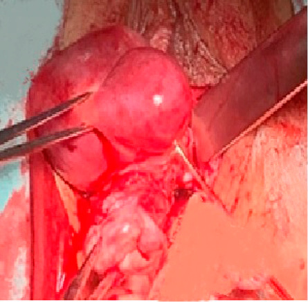
Introduction. Pelvic organ prolapse is increasingly spreading around the world. Therefore, surgical interventions in this pathology are becoming more frequent. Non-mesh surgery for pelvic organ prolapse is reported to have a higher incidence of disease recurrence, and polypropylene mesh implants appear to cause implant-associated complications, thereby limiting their application in clinical practice. Materials and methods. When a patient with an apical prolapse and high risk of postoperative complications sought medical care in the Clinic, the specialists decided to perform promontofixation using a titanium mesh implant. Results and discussion. The present paper describes a clinical case of surgical treatment of genital prolapse using a titanium mesh implant. Since the patient who was admitted for surgical treatment had a family history of genital prolapse, an application of non-mesh technologies implied a high probability of genital prolapse recurrence, as well as a high risk of the vaginal erosion after surgery. Therefore, a promontofixation was performed using a titanium mesh implant, and the 6-month follow-up period showed no postoperative complications. Conclusion. Analysis of postoperative complications demonstrated a considerable potential of using titanium mesh implants for the prevention of implant-associated complications.
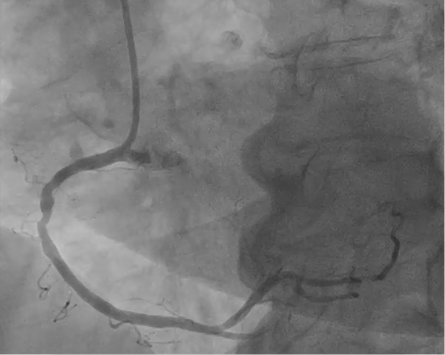
Introduction. An expanding number of indications for PCI in patients with coronary heart disease and severe concomitant pathology are accompanied by a growing number of patients with chronic renal failure. Contrast-induced nephropathy (CIN) is recognized as a severe complication, aggravating the course of the underlying disease, and, moreover, reducing the life expectancy of the patients. Modern intravascular imaging technologies are widely implemented in real clinical practice of endovascular surgery. In the context of increasing number of PCI performed in patients with severe concomitant pathology, the IVUS-guidance will improve the quality of stenting, and, importantly, lessen the risks of CIN due to the reduction in contrast volume. Materials and methods. The paper presents a clinical case of IVUSguided stenting of the right coronary artery without contrast agent in a patient with chronic kidney disease and the following diagnosis: “Coronary heart disease. Effort angina, class III (dyspnea as anginal equivalent). Balloon angioplasty and stenting of circumflex artery and LAD. Hyperlipidemia 2a. Atherosclerosis of the aorta, brachiocephalic and coronary arteries. Stage 3 hypertension. Controlled Hypertension. Level IV CVD risk. Type 2 diabetes mellitus. Target glycated hemoglobin is less than 7.5%. Grade 2 obesity, exogenous-constitutional. Renal microlithiasis. CKD stage 4 (GFR 29 ml/min/1.73m2). Cerebrovascular disease. Chronic cerebral ischemia.” Results and discussion. In the described clinical case, a complete myocardial revascularization was achieved using IVUS-guidance and minimal amount of contrast agent in a patient with severe CKD. The advantage of minimally invasive endovascular interventions in a complex category of patients, demonstrated by the case, implies expanded possibilities for providing high-tech care to patients with significant limitations in the use of contrast agents due to severe CKD with a high risk of CIN. Conclusion. Today, an increasing number of X-ray operating rooms in Russia are equipped with intravascular technologies, ensuring their wider use. The skills and knowledge in using IVUS imply rare application of contrast agents, thereby lessening the risk of CKD and, as a consequence, improving the prognosis of patients with reduced renal function and high risk of CKD.
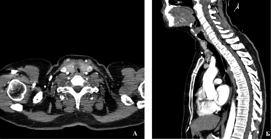
Introduction. Idiopathic cicatricial tracheal stenosis (ICTS) comprises a rare and dangerous disease of unknown etiology. Its morphological substrate consists in the formation of circular fibrotic stenosis, usually at the level of the cricoid cartilage and the first two С-shaped rings of the trachea. The first cases of the disease were described by Brandenburg only in 1972. Progression of the disease is slow, right up to severe respiratory failure and asphyxia. Treatment methods for idiopathic cicatricial tracheal stenosis include both endoscopic and open surgical techniques. However, endoscopic treatment fails to produce the desired effect. Materials and methods. The authors of the present paper performed tracheo-laryngeal resection as a radical method for treating idiopathic tracheal stenosis in a 54-year-old patient against the background of prolonged unsuccessful endoscopic treatment. The follow-up period accounted for 45 days. Examinations in the postoperative period showed complete epithelialization of the anastomosis line without inflammation. Results and discussion. Idiopathic cicatricial tracheal stenosis is considered to be a dangerous disease without significant causes of development. However, it is claimed to be a potentially curable disease, though its treatment is a difficult task. Some authors report satisfactory results of endoscopic treatment of ICTS, but most publications on this issue consider a single tracheo-laryngeal resection with anastomosis as the most optimal method of treatment, thereby correlating with the results of the present study. Conclusion. The present observation characterizes the peculiarity and possibility of radical treatment of ICTS, since the endoscopic method of treatment produces only a temporary effect. Small number of observations and lack of a coherent pathogenetic theory necessitate further research into this problem.

ISSN 2307-0501 (Online)


