


ORIGINAL ARTICLES

Introduction. Fracture of the proximal humerus is a common injury that accounts for up to 12 % of all bone fractures and up to 65 % of humeral fractures. 13 % to 16 % of fractures in this segment are multi-fragment with bone impression. This significantly complicates the task of internal fixation. This study aims to analyze the gender and age group distribution of patients with proximal humerus fractures, and morphological aspects of these injuries.
Materials and methods. This paper presents a retrospective analysis of the epidemiological and morphological parameters of patients treated surgically for proximal humerus injuries at the Department of Traumatology and Orthopedics of the State Regional Clinical Hospital of the Republic of Bashkortostan № 1 in the city of Oktyabrsky in 2010–2016. The total of 191 patients were included in the study, 121 (63.35 %) females and 70 (36.65 %) males.
Results and discussions. A signifi cant increase (more than three times) in the number of proximal humerus fractures was recorded in women in the 50–65 age group and in men in the 55–60 age group. The increase in the incidence of this type of injury does not exceed 22.4 % in comparison to the younger age groups. The increase in the number of injuries in question is undoubtedly due to changes in the bone metabolism in women. The analysis of character and morphology of fractures in women of older age groups indicates a greater prevalence of unstable injury of type 1.1.B and 1.1.C according to AO/ASIF classifi cation, which, again, is due to the demineralization of the segment determined by systemic metabolic abnormalities. The fi ndings indicate the need for perioperative monitoring of the bone metabolism parameters.
Conclusion.The morphology and types of fractures depend on a patient’s age and bone quality. A signifi cant increase in this pathology in women of perimenopausal age refl ects changes in bone metabolism.
The article presents the results of a retrospective study of the effectiveness of intestinal lavage with enteral saline solution for the treatment of dynamic intestinal obstruction in acute forms of pancreatitis and pancreonecrosis. The objective of this study is to improve treatment results in patients with intestinal paresis with various forms of acute pancreatitis with the use of intestinal lavage with enteral saline solution.
Materials and methods. The study included 81 patients, 56 (69.1 %) males and 25 (30.9 %) females, the age averaging at 59.3 ± 13.4 years. These patients were hospitalized at different time intervals counting from the onset of the disorder, ranging from 24 hours to 7 days. Patients were divided in two groups depending on hospitalisation prior to the first procedure of intestinal lavage providing there was no counterindications.
Results and discussion. Prokinetic effect of intestinal lavage in patients with gastrostasis and dynamic bowel obstruction help improve the quality of conservative treatment (up to 78.3 % in 1st group and 37.1 % in 2nd group); avoid open surgical procedures (up to 6.5 % in 1st group, up to 37.1 % in 2nd group), perform minimally invasive procedures to drain confined lesions (15.2 % of patients in 1st group, 42.9 % in 2nd group); eliminate manifestations of gastrostasis within 3 days following IL in both groups. The reduction/elimination of dynamic intestinal obstruction within 24 hours following IL (up to 73.8 % in 1st group, up to 97.1 % in 2nd group) made it possible to start early enteral nutrition within 48 hours (73.9 % in 1st group, up to 42.8 % in 2nd group).
Conclusions. Early use of intestinal lavage in the complex therapy of acute forms of pancreatitis is safe and effective. It reduces the number of purulent-septic complications, prevents the development of multiple organ failure, reduces the overall mortality, prepares the intestine for early enteral nutrition.

Introduction. Mantle cell lymphoma is a rare type of B-cell non-Hodgkin lymphoma. According to statistics the incidence of this disorder amounts to 2-3 per 100,000 people; this is about 6% of all non-Hodgkin lymphomas. It has been established that various molecular genetic characteristics of mantle cell lymphoma patients may present opportunities for a patient-specific approach to the disease prognosis and treatment strategy.
Materials and methods. The paper presents a retrospective analysis of 45 mantle cell lymphoma patients treated at the GAUZ RKOD of the Ministry of Healthcare from 2015 until now. The data used in the analysis included clinical examination, lab panels, PET CT, tumour and bone marrow biopsy specimen cytology, histology and immunohistochemistry. We analysed the epidemiological data, the patients’ clinical presentation characteristics, treatment approaches, immediate and long-term outcomes.
Results and discussion. We have established that the pathological process most frequently involves bone marrow (44%) and spleen (41%). The MIPI scores distribution was as follows: high in 14 (30%), medium in 20 (45%), low in 11 (41%). Ki67 was recorded at under 30% in four cases, in others it amounted to over 30%. In 2015–2017 patients were treated with the R-CHOP protocol with the following support with rituximab. PFS averaged at 20 months, 8 (17%) of patients remain in lasting remission (since 2015). In 2017 the R-BAC (high dose cytarabine for SCT candidates) and R-B (for the elderly and comorbid patients) became protocols of first line. Since 2018 eight patients have undergone auto-SCT (at the first late recurrence) as a treatment consolidation stage. As of now 13 patients have been examined in federal centres for del17p and the TP53 mutation.
Conclusion. We have demonstrated the need for and the option of treatment depending on the results of molecular genetic testing of mantle cell lymphomas.

Background. Our previous studies have shown that postnatally formed lymph nodes (PNFLN) can serve as a source of biological signals activating antitumour immune reactions and suppressing the spread of metastatic malignant cells.
Aim. To determine the expression of CD3, CD20, CD68 in native, sentinel and postnatally induced lymph nodes of the axillary zone in breast cancer.
Materials and methods. The study involved an analysis of digitalized images of the immunohistochemical expression of a fixed panel of antibodies CD3, CD20, CD68. The expression levels were assessed quantitatively by counting the expressed cells in each studied node for four main structural and functional zones.
Results and Discussion. The results of a comparative immunohistochemical study of native, sentinel and postnatally induced lymph nodes showed that the content of CD3, CD20, CD68 demonstrates fundamental differences in different lymph node structures.
Сonclusions
1. In postnatally induced lymph nodes, compared to native and sentinel lymph nodes, the distinct expression of antibodies to the main immunocompetent cells, which realize key immune responses in the lymph node, can indicate an increased functional status of the newly formed lymph nodes.
2. The study demonstrated a high level of antigenic stimulation of T and B lymphocytes in postnatally induced lymph nodes, as well as indicated a possible role of macrophage cells in the stimulation of neolymphogenesis and the formation of new lymph nodes.
3. The study provides the basis for further research into postnatal induced lymph nodes in cancer patients.
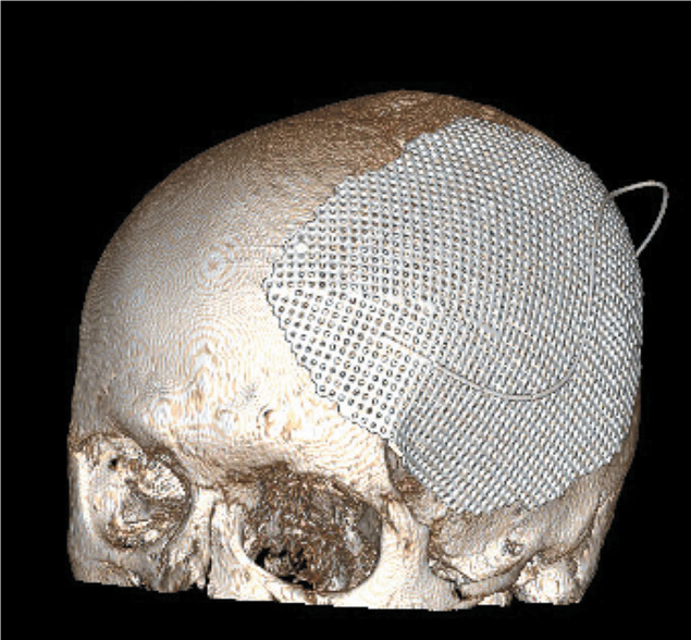
Introduction. A computed tomography (CT) scan of the entire body has become a standard practice for the diagnosis of patients with polytrauma in many trauma centres.
Purpose: evaluation of diagnostic potential of full-body CT for patients with polytrauma.
Materials and methods. In the period from January 2008 to December 2018, all hemodynamically stable patients (scoring 3 and above in the Abbreviated Injury Scale (AIS) and 15 and above in the Injury Severity Score (ISS)) admitted to the Main Republican Head Injury Centre CCH №21 in Ufa with injuries in two or more anatomical areas have undergone CT scanning of head, neck (including cervical spine), chest (including thoracic spine), abdominal cavity/pelvis (including lumbar spine).
Results and discussion. In the period from January 2008 to December 2018, 1498 CT examinations were conducted. Out of these examinations, 1368 and 143 cases were polytrauma — (on average 2 cases per week) and monotrauma (10 % of the study group) patients. Only 17 patients failed to complete the examination for polytrauma due to deteriorating status. All of these 17 had been returned to the intensive care unit without delay.
Conclusion. In a decade 1368 patients met the established criteria for an immediate CT scan for the diagnosis of polytrauma at a large hospital providing emergency healthcare. A broad range of significant injuries was diagnosed quickly, accurately and safely. These injuries included 31 cervical spine fractures and 56 pneumothoraxes not evidenced by conventional X-ray images.
REVIEWS
Regardless of such formidable figures medicine does not stand still; keeping up with the times, the science attempts to develop cutting edge methods of treating malignant tumours. As a result, the treatment of malignant neoplasms is continuing to improve. However, the number of side effects is also growing, thus requiring research attention. Therefore, the significance of the impact that oncological drugs have on a patient’s body is becoming more and more urgent for further discussion.
While current tumour treatment methods involving drugs such as tyrosine kinase inhibitors, anthracycline chemotherapy and immunotherapy protocols are effective for the treatment of various forms of cancer, these drugs affect the DNA replication process thus resulting in endothelial dysfunction and nonspecific immune response. This causes cardiotoxic side effects.
Cardiotoxicity, in its turn, is a notion that includes various adverse events involving the cardiovascular system of oncological patients receiving drug treatment. Cardiotoxicity may develop during treatment or following its completion.
It should be emphasized that the clinical significance of tertiary lymphoid structures ranges from a destructive to protective impact, which indicates the need for an improved understanding of the structure and case-specific function of these organs before conducting clinical targeting.
CLINICAL CASES

Introduction. Desmoid fibroma is a rare mesenchymal tumour developing from differentiated fibroblasts and excessive amounts of collagen fibres.
This paper presents a clinical case of removal of an anterior abdominal wall neoplasm — a rectus abdominis muscle desmoid tumour, with the following mesh implant reinforcement of the musculoaponeurotic layer.
Materials and methods. A 35 year old female patient Ch. was referred to the surgery department of “V nadezhnykh rukakh” hospital in November 2019, with complaints of a neoplasm in the anterior abdominal wall that was causing pain when touched, and dysuria. The patient’s clinical diagnosis was recorded as a neoplasm of the anterior abdominal wall.
Results and discussion. The patient underwent further examination and scheduled surgery. The neoplasm was removed completely leaving the surrounding healthy tissues, the anterior abdominal wall was reinforced with a mesh implant. The pathology results matched a rectus abdominis muscle desmoid tumour. Literature offers very little information on anterior abdominal wall neoplasms or procedures for their removal with simultaneous plastic mesh implantation. Cases such as this require further study.
Conclusion. A desmoid tumour of the rectus abdominis muscle can be radically removed; the procedure can be combined with the anterior abdominal wall plastic surgery with a mesh implant, making it possible to achieve a pronounced cosmetic effect regardless of the extensive surgical injury.
BRIEF COMMUNICATIONS

LETTER TO THE EDITORIAL OFFICE

ISSN 2307-0501 (Online)


